Research - Modern Phytomorphology ( 2021) Volume 15, Issue 3
Vascular anatomy and morphology of the flower of Hymenocallis latifolia (Mill.) Roem. (Amaryllidaceae)
O. Fishchuk*O. Fishchuk, Lesia Ukrainka Volyn National University, Pr. Voli. 13, Lutsk, 43025, Ukraine, Tel: 380663108107, Email: Fishchuk.Oksana@vnu.edu.ua
Received: 10-May-2021 Accepted: 20-Jul-2021 Published: 31-May-2021, DOI: 10.5281/zenodo.5801128
Abstract
The vascular anatomy and morphology structure of the flower of the Hymenocallis latifolia were studied. New morphological features in particular, vertical zonality of the gynoecium and vascular anatomy of the flower are not taken into account in the taxonomy of the family Amaryllidaceae. 10 flowers of Hymenocallis latifolia were sectioned using standard methods of Paraplast embedding and serial sectioning at 20 mkm thickness. Sections were stained with Safranin and Astra Blau and mounted in Eukitt. We investigated the presence of two vertical zones in the Hymenocallis latifolia gynoecium: symplicate and hemisymplicate. The micromorphology and vascular flower anatomy were described by using flowers transverse sections. Hymenocallis latifolia peduncle has 41 vascular bundles. The paired ventral bundles of the carpel supplied ovules. Above the locules, the ventral bundles of the carpel merge with the dorsal and septal vascular bundles, forming dorsal veins. Traces of dorsal vascular bundles of carpel are five-bundles, traces of septal bundles of carpel are three-bundles. Traces of outer tepals are thirteen-bundles, traces of inner tepals are nine-bundles. Traces of stamens are single-bundle. The new data helped to deepen the knowledge about the morphology and vascular anatomy features of Hymenocallis latifolia flowers and will help to compare the obtained morphological and anatomical features with the features studied earlier for members of the family Amaryllidaceae for further using them in taxonomy.
Keywords
Hymenocallis latifolia, ovary, vascular anatomy, vertical zones, vascular bundle
Introduction
Taxonomy of Amaryllidaceae family is based on modern molecular data (Chase et al. 2016; García et al. 2019), but there is a debate among researchers about the place of some genera in subfamilies and tribes, because micromorphological features and vascular anatomy features have not been used for family taxonomy before, while they are very important and unique. That’s why we examine Hymenocallis latifolia vascular anatomy and flower morphology. Some members of the family have been studied by us before (Fishchuk and Odintsova 2020; Fishchuk 2021).
The family Amaryllidaceae consists of 3 subfamilies and 14 tribes (Chase et al. 2016). The subfamily Amaryllidoideae includes genus Hymenocallis (tribe Hymenocallideae Small Andean), it has located inside the perigonium a corona–a special flower structure. The corona in Hymenocallis is formed by filament corona. The genus Hymenocallis has about 50 species distributed in the southeastern United States, the Antilles and from southern Mexico to Bolivia (Stevens 2020). Therefore, the aim of our work is to elucidate the features of the flower micromorphology and vascular anatomy and to identify its vertical zonality in H. latifolia.
Materials and Methods
Plant material was collected in the agricultural station of Lesya Ukrainka Volyn National University of Lutsk and fixed in 70% alcohol. Ten flower buds were dehydrated in t-butanol series (20%, 30%, 50%, 70 %, 100%-2 h each, the last one-24 h) and stored in 100% t-butanol and paraplast in the ratio 1:1. Infiltration was performed in Paraplast according to manufacturer’s instructions and R.P. Barykina(Barykina et al. 2004). Transverse and longitudinal sections of 20 μm thickness were obtained with manual rotary microtome (MPS-2 (USSR)) and stained in Safranin and Astra Blau. Slides were mounted in “Eukitt®” and images were obtained with an AMSCOPE 10MP digital camera attached to an AMSCOPE T490B- 10M (USA) microscope. For the morphological analysis, measurements were made on at least 15 fresh flowers. We used the concept of gynoecium vertical zonality by W. Leinfellner (Leinfellner 1950) to analyze the gynoecium’s internal structure. The height of the zones of gynoecium was measured according to the number of cross-sections. Vascular anatomy was studied on the series of flower cross sections.
Results
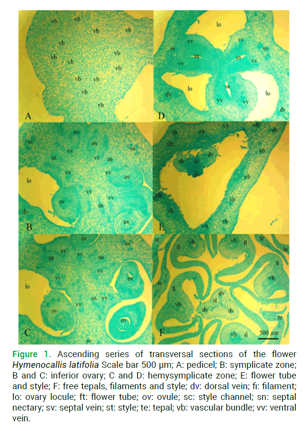
At the base of the peduncle H. latifolia has 41 vascular bundles (Fig.1A), a set of small and massive bundles, which are combined above to form a vascular cylinder. Slightly higher at the base of the receptacle deviated nine vascular bundles to the inner three tepals and thirteen vascular bundles to the outer three tepals (Figs.1E-1F) and six traces of stamens.

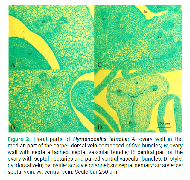
Figure 1:. Floral parts of Hymenocallis latifolia; А: ovary wall in the median part of the carpel, dorsal vein composed of five bundles; B: ovary wall with septa attached, septal vascular bundle; C: central part of the ovary with septal nectaries and paired ventral vascular bundles; D: style; dv: dorsal vein; ov: ovule; sc: style channel; sn: septal nectary; st: style; sv: septal vein; vv: ventral vein. Scale bar 250 μm.
At the level of the ovary base, three large vascular bundles are deflected–the dorsal bundles of the carpel (Figs.1B-1D) and three smaller–the septal bundles (Figs.1C-1D), and in the center remains a group of vascular bundles that diverge on both sides of the septal nectary–the roots of the ventral complex. Above the level of the ovule’s appearance, these bundles merge to form six massive vascular bundles–the ventral bundles of the carpel, which supplied ovules (Fig.1C).
Above the locules, the ventral bundles of the carpel (Fig.2C) merge with the dorsal and septal vascular bundles, forming dorsal veins (Fig.2D) that do not branch until the stigma. Traces of dorsal vascular bundles of carpel are five-bundles (Fig.2A), traces of septal bundles of carpel are three-bundles (Fig.1B). Traces of outer tepals are thirteen-bundles, traces of inner tepals are ninebundles. Traces of stamens are single-bundle.

Figure 2: Ascending series of transversal sections of the flower Hymenocallis latifolia Scale bar 500 μm; A: pedicel; B: symplicate zone; B and C: inferior ovary; C and D: hemysymplicate zone; E: flower tube and style; F: free tepals, filaments and style; dv: dorsal vein; fi: filament; lo: ovary locule; ft: flower tube; ov: ovule; sc: style channel; sn: septal nectary; sv: septal vein; st: style; te: tepal; vb: vascular bundle; vv: ventral vein.
In H. latifolia gynoecium we investigated the presence of following structural zones: fertile symplicate structural zone, height of which is about 100 μm (Fig.1B) and fertile hemisymplicate zone is about 2080 μm (Figs. 1C-1D). Septal nectaries appear in the hemisymplicate zone and open with nectary slits at the base of the style, the total height of the septal nectary is 2700 μm (Fig.1D). Style consists of asymplicate zone. The ovary roof is 620 microns.
Discussion
The flowers in the genus Hymenocallis have 2-10 ovules in each locule. Seeds are sometimes polyembryonic (Meerow and Snijman 1998). The karyotype of Hymenocallis littoralis (Tanee et al. 2018) and different species of the genus Hymenocallis were studied (Singh et al. 2017). In H. latifolia gynoecium presence symplicate and fertile hemisymplicate zones. In Hippeastrum striatum gynoecium were found synascidiate, symplicate and hemisymplicate vertical zones (Fishchuk 2021).
Morphologists are looking for new morphological features, such as internal micromorphological features and vascular anatomy flower features.
Conclusion
In the H. latifolia ovary available two vertical zones in the symplicate and hemisymplicate. The vascular system of the studied species characterized by the presence of multi-bundled traces of tepals, single-bundle stamens traces. The paired ventral bundles of the carpel supplied ovules. Traces of dorsal vascular bundles of carpel are five bundles, traces of septal bundles of carpel are three bundles. Flower morphology and vascular anatomy will help us to differentiate the studied species and their morphological and anatomy features from other members of the Amaryllidaceae family.
References
Barykina R.P., Veselova T.D., Deviatov A.G., Djalilova H.H., Iljina G.M., Chubatova N.V. (2004). Handbook of the botanical microtechniques. Moscow: Izd-vo MGU.
Chase M.W., Christenhusz M.J.M., Fay M.F., Byng J.W., Judd W.S., Soltis D.E., Stevens P.F. (2016). The angiosperm phylogeny group. An update of the angiosperm phylogeny group classification for the orders and families of flowering plants: APG IV. Botan J Linnean Soc 181: 1-20. https://doi.org/10.1111/boj.12385
Fishchuk O. (2021). Comparative flower morphology in Hippeastrum striatum (Lam.) H.E. Moore. (Amaryllidaceae). Ukr J Ecol 11: 273-278. https://doi.org/10.15421/2021_240
Fishchuk O.S., Odintsova A.V. (2020). Micromorphology and anatomy of the flowers of Galanthus nivalis and Leucojum vernum (Amaryllidaceae). Regul Mech Biosyst 11: 463-468. https://doi.org/10.15421/022071
Leinfellner W. (1950). Der Bauplan des syncarpen Gynoeceums. Österr Bot Zeitschr 97: 403-436.
Meerow A.W., Snijman D.A. (1998). Amaryllidaceae. In K. Kubitzki [ed.], Families and genera of vascular plants. Springer-Verlag 83-110. https://doi.org/10.1007/978-3-662-03533-7_11
García N., Meerow A.W., Arroyo-Leuenberger S., Oliveira R.S., Dutilh J.H., Soltis P.S., Judd W.S. (2019). Generic classification of Amaryllidaceae tribe Hippeastreae. Taxon 68: 425-612. https://doi.org/10.1002/tax.12062
Singh A., Misra S. (2017). Hymenocallis. Commercial Ornamental Crops: Traditional and Loose Flowers. Kruger Brentt Publishers UK LTD.
Stevens P.F. (2020). Angiosperm Phylogeny Website. http://www.mobot.org/MOBOT/research/APweb/
Tanee T., Sudmoon R., Siripiyasing P., Suwannakud K.S., Monkheang P., Chaveerach A. (2018). New Karyotype Information of Hymenocallis littoralis, Amaryllidaceae. Cytologia 83: 437-440. https://doi.org/10.1508/cytologia.83.437