Research - Modern Phytomorphology ( 2022) Volume 16, Issue 2
Anatomical structure of seed coat in Pombalia bigibbosa and P. calceolaria from Argentina (Violaceae)
Micaela-Noemí Seo1* and Maia Fradkin2,32Faculty of Agricultural Sciences, University of Lomas de Zamora, Camino de Cintura 1055, Lavallol (1836), Province of Buenos Aires, Argentina
3National Council for Scientific and Technical Research, Argentina
Micaela-Noemí Seo, Institute of Health Sciences, Arturo Jauretche National University, Av. Calchaqui 6200, Florencio Varela (1888), Province of Buenos Aires, Argentina, Email: micaseo@gmail.com
Received: 24-Feb-2022, Manuscript No. mp-22-55459 ; Accepted: 25-May-2022, Pre QC No. mp-22-55459 (PQ); Editor assigned: 25-Feb-2022, Pre QC No. mp-22-55459 (PQ); Reviewed: 25-May-2022, QC No. mp-22-55459 (Q); Revised: 25-May-2022, Manuscript No. mp-22-55459 (R); Published: 02-Jun-2022, DOI: 10.5281/zenodo.7735589
Abstract
In this study, the anatomical structure of seed-coat of Pombalia bigibbosa (A. St.-Hil.) Paula-Souza and P. calceolaria (L.) Paula- Souza from Argentina was studied for the first time in the Violaceae. The anatomy of seed-coat revealed the presence of three layers in testa derived from outer integument in both species however the presence of layers in tegmen was different between the analyzed species, P. bigibbosa presented two layers meanwhile P. calceolaria exhibited three layers derived from the inner integument in the mature seed-coat. The presence of a conspicuous elaiosome with oily drops was observed in both species, this structure had been observed in the genus Viola and it would be related with the attraction and dispersion mediated by ants in these species of the genus Pombalia in nature. In this sense, the myrmecochory in these violets species explains the multiple evolutionary origins in the angiosperm plants and is proposed like an example of convergent evolution.
Keywords
Pombalia, elaiosome, myrmecochory, seed-coat
Introduction
troduction In a molecular analysis of the family Violaceae Batch., Wahlert et al. (2014) revealed the polyphyletic origin of genus Hybanthus Jacq., and according to this analysis, most of the species of this genus were transferred into the new genus Pombalia Vand. (Paula-Souza & Ballard 2014). The genus Pombalia Vand. comprises about 40 mostly of herbs and shrubs is distributed throughout most temperate regions of Latin America and South America, in Argentina includes 15 native species (Seo et al 2017).
In the family Violaceae, there are anatomical studies of seeds covering of the genus Viola L. (Shahrestani et al. 2014; Culver & Beattie 1978). According to the traditional description in Violaceae published by Corner (1976), the seeds of Pombalia are exostegmatic in relation of the position of the mechanical layer, present a ovoid to globose shape and possess a conspicuous linear raphe and a exostomal elaiosome. The anatomical studies related to Hybanthus species is based on the embryology of Ionidium suffruticosum (L.) Roth ex Schult., a synonym of Hybanthus suffruticosus (L.) Baill. published by Raju (1958) and Singh (1961).
Viola presents an elaiosome which originates as an outgrowth of the micropyle or the raphe base.
Seed dispersal by ants or myrmecochory has added greatly to knowledge on the ecology in the ant–plant mutualisms in flowering plants (Lengyel et al. 2010). The genus Viola presents an elaiosome which originates as an outgrowth of the micropyle or the raphe base (Corner 1976), this structure is a lipid-rich appendage and the function is to attract ants and elicit the transport of the seed to the nest by the ants (Lengyel et al. 2010; Beattie and Lyons 1975; Culver and Beattie 1978) and function as rewards for ants because contains reserves with lipids or proteins (Lisci & Pacini, 1997; Lengyel et al. 2010). The presence of elaiosome was mentioned in the genus Hybanthus (Lengyel et al. 2010; Seo 2010).
This paper provides a detailed description of anatomical features of seed-coat of P. bigibbosa (A. St.-Hil.) Paula-Souza and P. calceolaria (L.) Paula-Souza in order to analyze the layers of tegmen and testa. To determine the presence of myrmecochory, the anatomical feature of elaiosome is observed to evaluate the possible dispersion by ants in nature.
Materials and Methods
Plants with seeds were collected from natural habitats of northern Argentina and each specimen was given a voucher and a collector’s number and deposited at Buenos Aires University Herbarium (BAFC). The collection data for the examined specimens are given in Tab. 1.
Table 1. List of localities and voucher of the collected seeds of P. bigibbosa and P. calceolaria in Argentine.
| Species
|
Procedence and voucher number
|
|---|---|
| P. bigibbosa | Misiones. Dpto. Iguazú, Pto. Iguazú, PN. Cataratas del Iguazú, sendero Macuco, sendero Macuco. Seo 7 (BAFC). |
| P. bigibbosa | Misiones. Dpto. Iguazú, Pto. Iguazú, PN. Cataratas del Iguazú, sendero Macuco, a 500 m. del CIES. Seo 19 y 27 (BAFC).
|
| P. bigibbosa | Misiones. Dpto. Iguazú, Pto. Iguazú, PN. Cataratas del Iguazú, sendero Macuco, a 1800 m. del CIES. Seo 23 (BAFC). |
| P. bigibbosa | Misiones, Dpto. Iguazú, PN. Cataratas del Iguazú, en la entrada del CIES, en los jardines alrededores del CIES. Seo 47 (BAFC). |
| P. calceolaria | Corrientes. Dpto. Ituzaingó, barrio Mil viviendas Yaciretá. En camping Municipal Soro. Seo 1 (BAFC). |
| P. calceolaria | Corrientes. Dpto. Ituzaingó, barrio Mil viviendas Yaciretá. Zona alta de lomada, camping Municipal Soro. Seo 16 y 17 (BAFC). |
| P. calceolaria | Corrientes, Dpto. Ituzaingó, Bo. Mil Viviendas, en el Balneario Soro. Seo 59 (BAFC). |
Collected seeds were fixed in 70% alcohol for 3 days. Transverse sections of lateral stems and leaves were made manually using commercial razor blades. The cross sections were stained with Toluidine Blue (O-Brien et al. 1964). Seed-coat layers was classified using current terminology described by Singh (1961) and the nomenclature used in the seed description was that defined by Corner (1976).
According to Singh (1961), the seed coat develops from the Inner Integument (II) and the Outer Integument (OI). The testa is formed from the three layers of the OI, the exotesta or Outer Testa (TSO) is an epidermis cuticularised and its cells have cellulose thickening of lamellate type. The cells of the middle testa (TSM) are thick-wailed and elongated at right angles. And the presence of calcium oxalate crystals is from the inner OI (TSI) which remains closely appressed to the sclerenchyma. In the tegmen, the Mechanical Layer (ML) or sclerenchyma derived from the outer II. The cells of the middle layer (TGM) of the II practically disappear, while those of the inner II (TGI) after thickening form the lining layer of the seedcoat.
Results
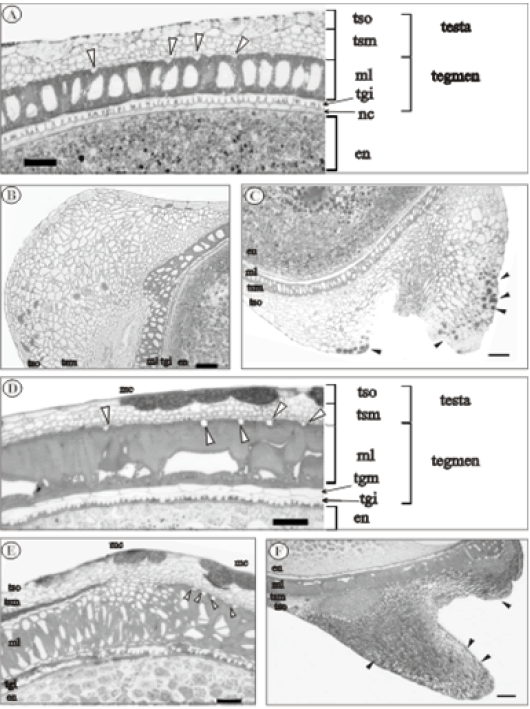
The seed coat development of the analyzed species of Pombalia resembles the embryology of I. suffruticosum (Raju 1958; Singh 1961). Thus, according to the present investigation the three layers of the OI take part in the formation of the seed-coat and are in agreement with the presence of calcium oxalate crystal near to the ML, however differs in minor details in the layers of inner II and this differences are discussed in the present analysis of P. bigibbosa (Figs. 1A-1C) and P. calceolaria (Figs. 1D-1F).
Figure 1: Pombalia bigibbossa (A,B and C). A: T.S. of seed coat with five layers of testa and tegmen (white triangle pointing to crystal); B: L.S. in chalazal region; C: L.S. with the elaiosome (black triangle pointing to oily drop); Pombalia calceolaria (D, E and F). D: T.S. of the seed coat with six layers of testa and tegmen (white triangle pointing to crystal); E: L.S. in chalazal region; F: L.S. with the elaiosome (black triangle pointing to oily content). (En: endosperm, nc: nucellus, mc: mucilage, ml: mechanical layer, tsm: middle testa, tso: outer testa, tgi: inner tegmen, tgm: middle tegmen. Scale bars: 10 μm (A-D), 200 μm (B, C, E, F).
The size of epidermal cells in each layer of the seed coat varies between the region of the seed, in the chalazal area (Fig. 1B and Fig. 1E) are longer than the lateral sides of the seed (Fig. 1A and Fig. 1D) in both species.
In transverse sections of the seed in both species of Pombalia (Fig. 1A and Fig. 1D) in the TSO there is cutinized epidermis with elongated cells and possesses mucilagous cells in P. calceolaria (Fig. 1D). Inside the TSM is formed by three to four layers of small and isodiametric cells, thick-walled and elongate at right angles to the cells of the mechanical layer. The oxalate crystals is observed in both species, the crystals are triangular to rectangular in P. bigibbosa (Fig. 1A) and quadrangular in P. calceolaria (Fig. 1D).
In the tegmen of both species there is also a single layer of exotegmic sclereids in the ML however the shape of the thick wall are quite different. P. bigibbosa presents braquysclereids (Fig. 1A) with massive thickened walls with a central pith however P. calceolaria present cells similar in shape similar to a osteosclereids the thickened is irregular to the anticlinal walls (Fig. 1D). Inside the inner epidermis of tegmen is covered by two layers of cells in both species (Fig. 1A and Fig. 1D): one layer presents small and isodiametric cells with thick-walled and the cells of other layer are rectangular with thin walls and without content in the interior. The disposition of these two layers differs between species, in P. bigibbosa the cells of outer of TGI is thick walled and the inner is thin walled (Fig. 1A), in P. calceolaria the cells thick walled are in the inner tegmen and the thin walls in the outer tegmen (Fig. 1D). According to Singh (1961), the TGM disappear and TGI persists in a mature seed, and based on the position of these layers, in P. bigibbosa the layer inside the TGI (Fig. 1A) corresponds to nucellus, which persist near the endosperm, and in P. calceolaria the layer of rectangular cells with thin walls corresponds to the TGM (Fig. 1D) which persist in the mature seed.
In the chalazal region of the seed, the layers of parenchymatous cells varies between specie located between the TSO and ML, P. bigibbosa presents more than twenty layers of parenchyma cells (Fig. 1B) and P. calceolaria exhibits four to six layers of parenchyma in TSM (Fig. 1E).
A longitudinal elaiosome was situated in the micropilar part of the seed in both species (Fig. 1C and Fig. 1F). In the transverse section of P. bigibbosa the elaiosome exhibit a triangle shape with an external layer of TSO and in the TSM twenty to thirty layers of small and isodiametric cells with thin walls, elongated to the right angles to the cells, some cells of TSO and testa presents oily drops in the outer elaiosome structure (Fig. 1C). In the transverse section of P. calceolaria the elaiosome is elongated like a tubular shape with an external layer of TSO and in the TSM near to twenty layers of small and isodiametric cells with thin walls, some cells of TSO presents oily drops (Fig. 1 F).
Discussion
Knowledge regarding the anatomical studies of seed coat in the genus Pombalia has not been studied, the first comprehensive research of this genus in Argentina. The seed coat development of the analyzed species of Pombalia resembles the embryology of I. suffruticosum (Singh 1961; Raju 1958), these authors concluded that all layers of the OI take part in the formation of the seed-coat (Singh 1961). Seed coat in the two species of Pombalia derived from II and OI however the number of layers in the inner tegmen varies between species, P. calceolaria presents six layers from both integuments, similar to the observations of Raju (1958) however P. bigibbosa presents five layers, TGM disappear in mature seed, similar to the description of Singh (1961). The presence of oxalate crystals and a single ML is similar in both species. The two species further differ in shape of the sclereids would provide additional taxonomic in the delimitation of species with fruits, which is one of the major unresolved problems in the specific identification of Hybanthus and Pombalia (Seo 2010).
In both species in the region of the microphyla the elaiosome is conspicuous and in some cells of the testa presents oily drops, similar to the anatomy of the elaiosome previously observed in Viola (Culver & Beattie 1978; Singh 1961). In this sense, the presence of oil or lipid content in both species of Pombalia would suggest to be more attractive to the ants (Lengyel et al. 2010; Lisci & Pacini 1997) and the myrmecochory would be the dispersion of these seeds in the nature. Lengyel et al. (2010) suggested that the low costs of producing an elaiosome and the consistent selective benefits of myrmecochory, explain the numerous evolutionary origins in the angiosperm plants and proposed like an example of convergent evolution in biology.
Conclusions
In the light of these findings, the anatomical analysis of the seed coat revealed and would provide information with high significance in the embryology of this genus Pombalia in order to improve the knowledge in Violaceae. However, further studies will be necessary to complete and the anatomy and embryology of the seed coat of these species has not been analyzed in the most species of Pombalia and will be essential to understand the interaction between ants and elaiosome in myrmecochory of seed dispersion in natural habits.
Acknowledgements
The authors thank to the curators of CTES and SI and also to the National Parks Administration of Argentina. This study was supported by CONICET and IAPT Research Grants 2007.
References
Beattie A.J., Lyons N. (1975). Seed dispersal in Viola (Violaceae): adaptations and strategies. Amer J Botany 62: 714-722.
Corner E.J.H. (1976). The Seeds of Dicotyledons: Volume 1. Cambridge University Press pp: 311.
Culver D.C., Beattie A.J. (1978). Myrmecochory in Viola: dynamics of seed-ant interactions in some West Virginia species. J Ecol 1: 53-72.
Lengyel S., Gove A.D., Latimer A.M., Majer J.D., Dunn R.R. (2010). Convergent evolution of seed dispersal by ants, and phylogeny and biogeography in flowering plants: a global survey. Perspect Plant Ecol Evol Systematics 12: 43-55.
Lisci M., Bianchini M., Pacini E. (1996). Structure and function of the elaiosome in some angiosperm species. Flora 191: 131-141.
O'Brien T., Feder N., McCully M.E. (1964). Polychromatic staining of plant cell walls by toluidine blue. O. Protoplasma 59: 368-373.
Paula-Souza J.D., Ballard Jr H.E. (2014). Re-establishment of the name Pombalia, and new combinations from the polyphyletic Hybanthus (Violaceae). Phytotaxa 183: 1-15.
Raju M.V.S. (1958). Seed development and fruit dehiscence in Ionidium suffruticosum Ging. Phytomorphology 8: 218-224.
Mohammadi Shahrestani M., Saeidi Mehrvarz S., Marcussen T., Yousefi N. (2014). Taxonomy and comparative anatomical studies of Viola sect. Sclerosium (Violaceae) in Iran. Acta botanica gallica 161: 343-353.
Singh D. (1963). Structure and Development of Ovule and Seed of Viola Tricolor L. and Ionidium Suffruticosum Ging. J Indian Bot Soc 42: 448-462.
Seo M.N. (2010). Seed coat micromorphology of South American species of Hybanthus (Violaceae). Nordic J Botany 28: 366-370.
Wahlert G.A., Marcussen T., de Paula-Souza J., Feng M., Ballard H.E. (2014). A phylogeny of the Violaceae (Malpighiales) inferred from plastid DNA sequences: implications for generic diversity and intrafamilial classification. Systematic Bot 39: 239-252.
